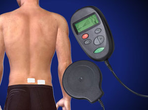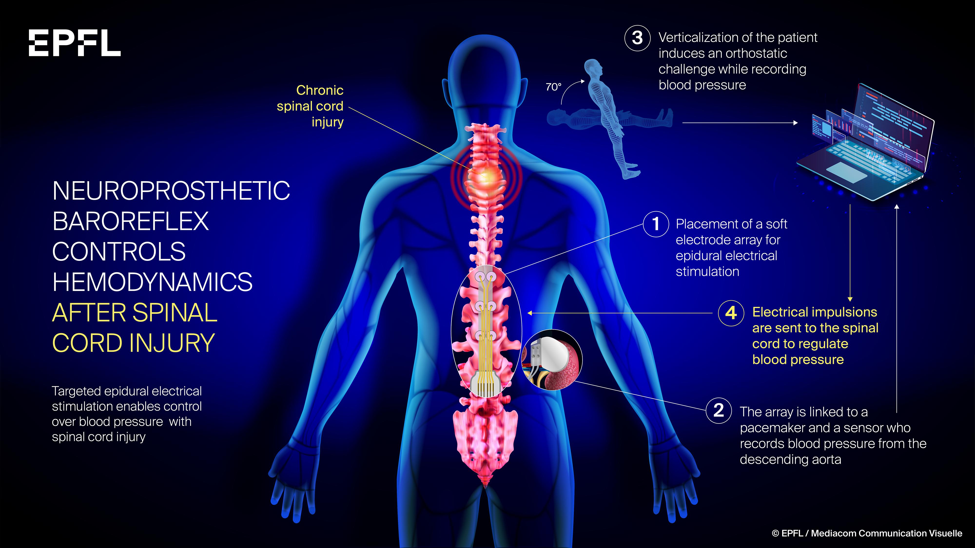


The site on the back where the leads and electrodes were inserted is usually uncomfortable for a few days.
SPINAL CORD STIMULATOR IMPLANT PICTURES HOW TO
See How to Prepare Psychologically for Back Surgery Baseline leg pain mean of 8, dropped to 3.2, and baseline low back pain mean of 7.5, dropped to 4.2 at 12 months. If the pain is not relieved, the doctor should be contacted right away so the device can be reprogrammed. Tests paddle-shaped SCS lead efficacy in capturing both back and leg pain. Multi-Lead Trialing Cable for Spinal Cord Stimulation.
SPINAL CORD STIMULATOR IMPLANT PICTURES TRIAL
The wire connecting to the external neurostimulator is taped to the person’s back during the trial to hold it in place. The leads are connected to an exterior pulse transmitter that the patient wears on a belt.Once the patient has reported on the pain-relief coverage, the patient is again sedated. Devices using newer technology generally avoid the tingling sensation and a wake-up test may not be needed.) Each electrode affects pain in a different area, so doctor-patient communication is crucial in making sure the doctor has adjusted the location of the electrical contacts to cover all the areas in pain. (If a low-frequency system is used, the goal will be to cover all painful areas with a slight tingling sensation known as paresthesia. The patient is awakened to provide feedback on the specific areas where pain is relieved by the stimulation, and where pain relief is still required.See Introduction to Diagnostic Studies for Back and Neck Pain In some cases, a small incision may be needed to insert the needle. It has one or two leads that attach to your spinal. The needle contains thin, insulated wires, called leads, with electrical contacts attached. A spinal cord stimulator is something your Alliance Spine Associates, LLC doctor can implant in your body. Guided by fluoroscopy (a type of X-ray), the doctor inserts a hollow needle into the area around the spinal canal called the epidural space.Local anesthesia is applied to the injection site and sedation may be provided.Procedures vary somewhat with the stimulation device used, but these are the typical steps. The trial period procedure is usually performed in a doctor’s office or a surgical center.


 0 kommentar(er)
